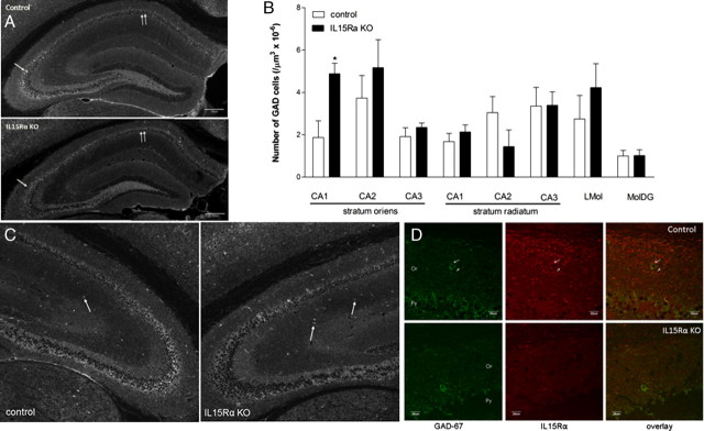
Figure 4.
IHC revealed a region-specific distribution of GAD67 immunoreactivity that showed an increase in the number of interneurons in the stratum oriens of the CA1 region in the IL15Rα KO mice. n = 3/group. A, In the control mice, two types of GAD-67 immunostaining were seen. This includes the soma of interneurons dispersed in the dendritic layer, and synaptic terminals in the granular cell layers in CA and DG regions (arrows). In the KO mice, there seemed to be a subtle increase of the number of scattered immunopositive neurons but a reduction in the staining intensity of the synaptic terminals. B, Cell counts showed that the KO mice had significantly more GAD-67 (+) neurons only in the stratum oriens of the CA1 region. *p < 0.05. C, Representative images showing increased number of GAD-67 (+) interneurons in a KO mouse (right panel) in comparison with the control mouse (left panel). D, Colocalization of IL15Rα with GAD-67 in the control mice was seen in some interneurons and cellular processes of the stratum oriens of the CA1 region (Or, arrows), and to a lesser extent in the pyramidal cell layer (Pr). The immunoreactivity of IL15Rα was absent in the Or region of the KO mouse, but the staining for GAD-67 persisted.

