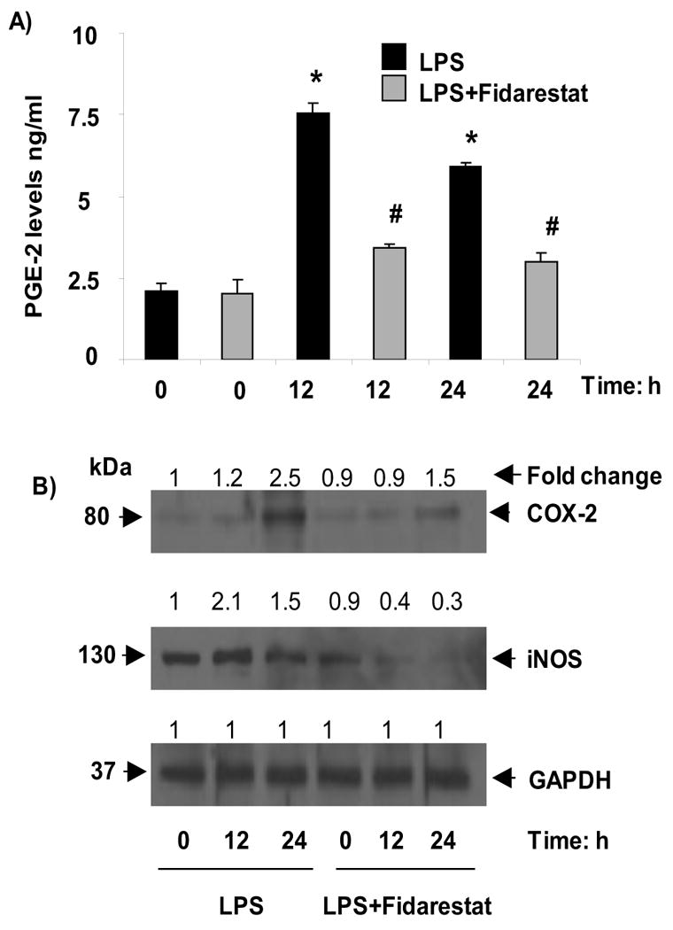
Fig.6. AR inhibition prevents LPS-induced inflammatory markers.

Growth-arrested RAW264.7 cells were incubated with and without 2μM fidarestat for 24 h and then treated with 1μg/ml LPS for 0, 12 and 24 h. A) PGE-2 was measured in cell culture medium and B) whole cell lysates were used for Westren blot analysis and probed with COX-2, iNOS and GAPDH antibodies. Band intensities were measured by densitometric analysis to determine the fold change. All the data are expressed as Mean ± SEM (N = 4). *P < 0.001 as compared to control cells, #P < 0.001 compared to LPS-treated cells
