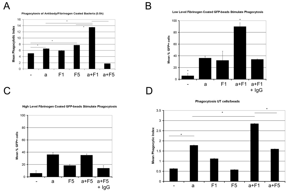
Figure 2. Different levels of fibrinogen present during the antibody-coating of particles produces a bimodal phagocytosis response.
Low levels of fibrinogen (1 mg/ml fibrinogen, F1) present during the coating of bacteria with antibodies (a), result in enhanced phagocytosis of these bacteria by macrophages when challenged with these bacteria in serum-free media(A). When coating sterile GFP-coated polystyrene beads with combinations of anti-GFP IgG and fibrinogen, the same effect was observed, and presence of excess non-specific IgG during the coating progress inhibited this response in macrophages (B). Similar as observed with bacteria, high levels of fibrinogen (5 mg/ml, F5) during the coating of GFP-beads with anti-GFP IgG, did not produce any subsequent enhanced phagocytosis in macrophages(C). When repeating these experiments with undifferentiated THP-1 cells that lack beta2 integrin receptors for fibrinogen, different levels of fibrinogen present during the coating process of GFP-coated beads with anti-GFP antibodies still produced different phagocytosis responses (D). Experimental Details: GFP-expressing E.coli or GFP-coated polystyrene beads were coated with antibody and/or fibrinogen at various concentrations for 1 hour at 25°C. Bacteria/beads were washed thoroughly to remove free fibrinogen and antibody, and then applied to macrophage or THP-1 cultures in serum-free media. A) Phagocytic indices after 0.5 hour in 40–70 macrophages, B, C) Mean % of cells containing more than 1 GFP bead after 0.5 hours in 130–180 macrophages. In B) particles were coated with anti-GFP antibody and/or 1 mg/ml Fibrinogen, while in C) particles were coated with anti-GFP antibody and/or 5 mg/ml Fibrinogen. D) Mean phagocytosis of anti-GFP IgG or fibrinogen coated GFP-beads in undifferentiated THP-1 cells after 1 hour. For all figures: a=antibody 1 µg/ml, F1=1 mg/ml fibrinogen, F5=5 mg/ml fibrinogen, IgG=5000-fold excess unspecific IgG added. Beads were incubated under these conditions for 1 hour and washed three times before the phagocytosis assay to remove unbound fibrinogen or antibody. All experiments were repeated three times producing consistent results.

