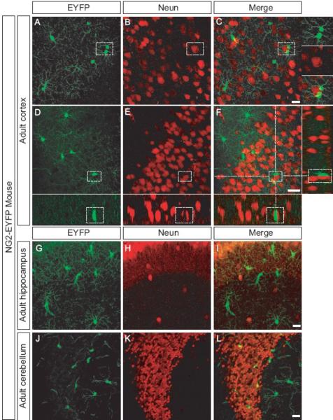
Figure 3. Intimate contact between neurons and NG2 cells in the CNS.
Confocal image scan of cortex, hippocampus, and cerebellum of adult mice expressing EYFP (A,D,G, J ) stained with an antibody that recognizes Neun (B, E, H, K). Merged images (C, F, I, L) shows no overlap, but close association between EYFP+ cell and Neun + neurons. Inserts at high magnification show EYFP+ cells close to Neun+ neurons.
Scale bars = 20 μm

