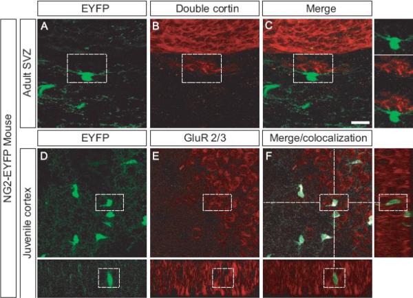
Figure 4. Intimate contact between neurons and NG2 cells in the SVZ and expression of GluR 2/3 by NG2 cells in vivo.
Confocal image scan of the SVZ of adult mice expressing EYFP (A), stained with an antibody that recognizes Doublecortin (B). Merged images (C) show no overlap but close association between EYFP+ cells and Doublecortin+ neurons. Hogh magnification inserts show EYFP+ cells very close to Doublecortin + neurons. EYFP+ cells (D) in the juvenile cortex stain with an antibody recognising GluR2/3 (E). Merge and co-localization analysis shows expression of the GluR2/3 on the processes and cell body of the EYFP+ cells.
Scale bars = 20 μm

