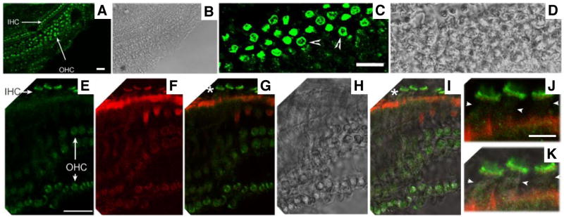
Fig. 2. CFTR protein expression in the cochlea.
CFTR is stained with anti-CFTR (green color, H182, Santa Cruz). Actin is stained with Texas Red-X phalloidin (red color, Molecular Probes). A–B. Immunofluorescence (A) and the corresponding phase-contrast images (B) of a whole-mount cochlea taken at a low magnification (20× objective). C–D. Immunofluorescence (C) and the corresponding phase-contrast images of a whole-mount cochlea show a cross section of OHCs. The lateral membrane of OHCs is indicated by arrowheads. E–I. Immunofluorescence (E and F) and the corresponding phase-contrast images (H) of a whole-mount cochlea. Mechanical manipulation was used to adjust cochlear samples, allowing some hair cells to lie flat on the slide. As a result, a quasi side-sectional image of IHCs was obtained. G. Superimposed images from E (green) and F (red). I. Superimposed images from E, F and H (phase contrast). J–K. These images correspond to the locations marked with “*” and are given at higher magnification for better examination of the lateral membrane of IHCs (arrowheads). Bar length: 20 μm (A, C, and E), 7 μm (J).

