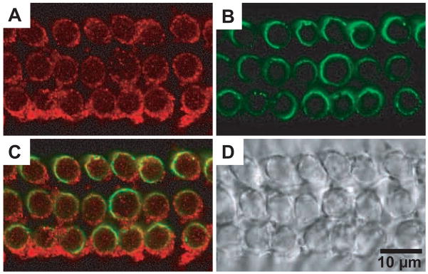
Fig. 4. Co-immunostaining of CFTR and prestin in OHCs.
CFTR and prestin were co-stained with anti-CFTR (red, M-15, Santa Cruz) and anti-mPres (green), respectively. A. CFTR staining. B. prestin staining. C. Superimposed image from A and B. D. Phase-contrast image. The ring-like staining pattern for CFTR was not observed in OHCs derived from prestin-KO mice (not shown). The results are consistent with the observations in the preceding figures (Fig. 2 and 3).

