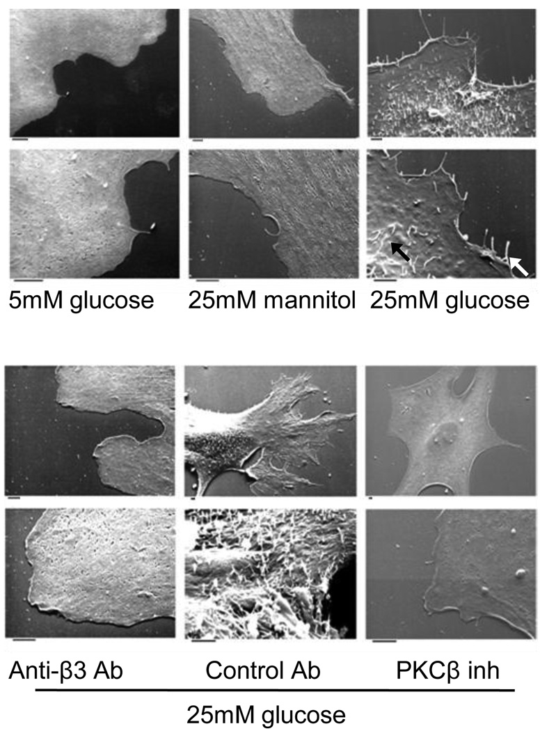
Figure 2. Hyperglycemia triggers dorsal and lateral filopodial-like projections in SMCs.
Electron micrographs of adherent SMCs cultured in euglycemic (5 mM) or hyperglycemic (25 mM) conditions with the indicated inhibitors. Inhibitors used were as follows: PKCβ(3-(1-(3-Imidazol-1-ylpropyl)-1H-indol-3-yl)-4-anilino-1H-pyrrole-2,5-dione, 5nM), anti-β3 antibody (10µg/ml), control antibody (10µg/ml). The photomicrographs are representative of results obtained in five experiments. The white arrow head (→) points to the lateral filapodia and black arrow head(→) points to the dorsal filapodia. Bar denotes 2µm.

