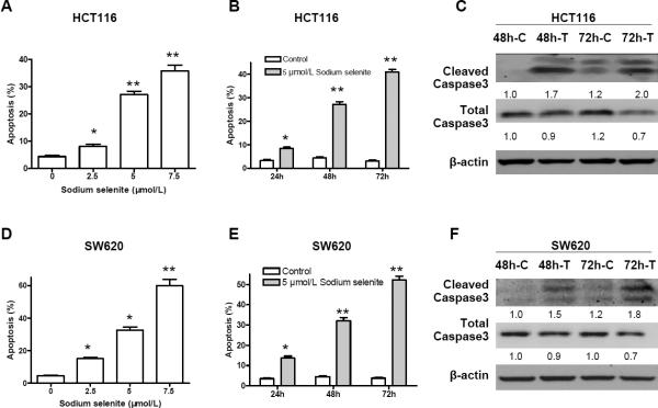
Figure 3.
Sodium selenite promoted apoptosis in colon cancer lines HCT116 and SW620. A and D, apoptosis induction by sodium selenite was in a dosage dependent manner (48h); B and E, apoptosis induction by sodium selenite was time-dependent (5μmol/L); C and F, increase of cleaved Caspase 3, a biomarker of apoptosis, was observed in both colon cancer cell lines after 48 h and 72 h treatment of 5 μmol/L of sodium selenite, while the total caspases 3 was slightly reduced. “C” referred as “control”, and “T” referred as “sodium selenite treatment”. β-actin was used as loading control. The image signal was quantified and normalized to β- actin. The ratio was presented. These experiments were triplicated independently. (* p<0.05, ** p<0.01, compared to control, respectively).

