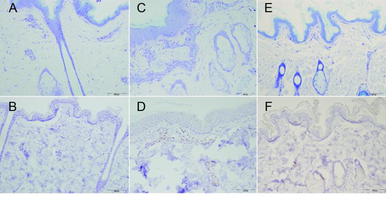
Figure 2. Phenotypic characterization of skin biopsies.
Photomicrographs are from a macaque with normal skin (A and B) a macaque with moderate alopecia (C and D) and a Cynomolgus macaque (E and F) showing increased mast cells in the alopecic Rhesus macaque (C) compared with normal Rhesus and Cynomolgus skin (A and E) and increased CD3+ lymphocytes (D) compared with normal Rhesus and Cynomolgus skin (B and F). Top row, Toluidine blue staining mast cell granules purple and bottom row CD3 immunostain using avidin-biotin-peroxidase method with DAB counterstain.

