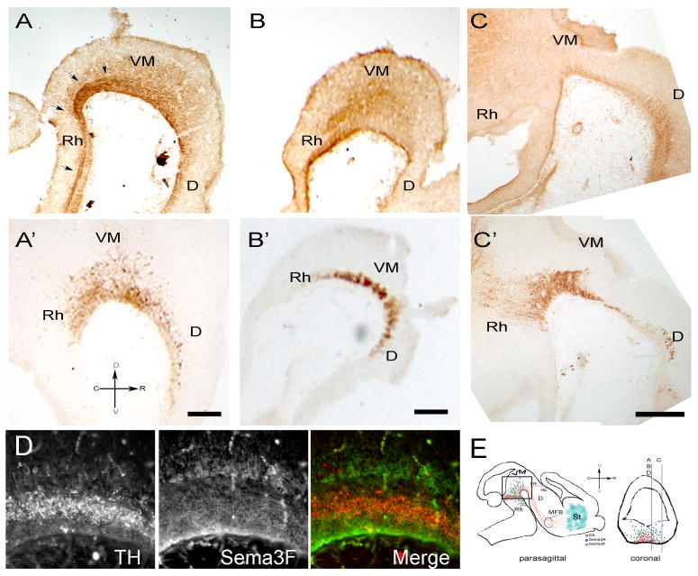
Figure 1. Sema3A and Sema3F are strongly expressed in the ventral mesencephalon of E13.5 VM rat embryos.
E13.5 rat embryos were fixed and 40μm adjacent sagittal sections were stained with antibodies against Sema3A (A), Sema3F (B, C) or TH (A′, B′, C′). In the VM (A), Sema3A is found as an increasing gradient extending from the floor of the ventricle to the pial surface. Medial sections showed a dense track of axons originating in the rostral mesencephalon, presumably the medial longitudinal fasciculus, which was strongly stained for Sema3A (arrows). Sema3F is expressed in a region that appears to wrap the DA neurons at their dorsal, ventral and caudal borders while presenting an ostensibly decreasing gradient in the rostral direction. The caging pattern dissipates in lateral regions of the VM (C). (D) Double staining with TH (red) and Sema3F (green) antibodies confirms the caging distribution of Sema3F. (E) Schematic diagram of the sectioning strategy used along these studies. Stained sections were cut parasagittally within 500 μm from the midline as represented in a coronal section for panels A, B, C and D. All pictures were taken in the region depicted by the frame in the sagittal cut. Spatial orientation is represented by a cross where C, R, D, and V represent caudal, rostral, dorsal and ventral positions. Scale bars: 100 μm. D, Diencephalon; Rh, Rhomboencephalon; FR, fasciculus retroflexus; Hb, habenula.

