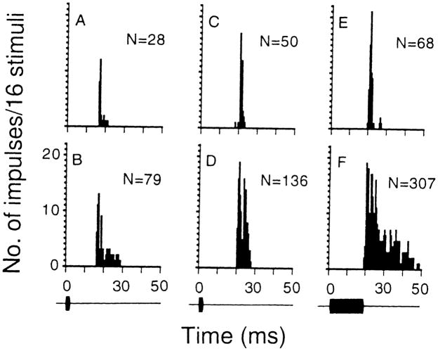
Fig. 2.
A–F PST histograms showing the discharge patterns of two inferior collicular neurons obtained with 80-dB SPL sounds before and after bicuculline application to their recording sites. The discharge patterns of A–D were obtained with 3-ms sounds and those in E, F were obtained with 19-ms sounds. The discharge pattern of one collicular neuron (BF: 43.5 kHz, MT: 22 dB SPL) changed from phasic responder (A) into phasic burster (B) after bicuculline application. The discharge pattern of another neuron (BF: 35.1 kHz, MT: 47 dB SPL) changed from phasic responder (C, E) into tonic responder (D, F) after bicuculline application such that this neuron discharged impulses with a duration longer than the presented pulses. N: total number of impulses in the histogram. Bin width: 500 μs, sampling period: 300 ms. To highlight the change in discharge pattern, the sampling period beyond 50 ms is not shown

