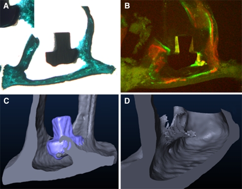
FIG. 3.
Osseointegration of a collagen I+decorin+bone morphogenic protein 4-coated titanium footplate anchor. A The histological section shows the implant embedded at its edge with a bone isodense brace-shaped formation. B The fluorochrome sequence labeling clarifies the new bone a formed between days 21 and 28 after implantation. The three-dimensional reconstruction of the specimen (C) illustrates the brace-like shape of the bone formation. D Again, the computed implant elimination brings out the greater extend of osseointegration along the implants edge. A view from the rear is illustrated for better visualization of the newly formed bone.

