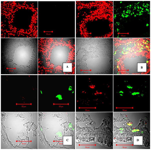
Fig. 1.
Staining for GFP in paraffin and cryosections in the mouse lung at 6–8 weeks following GFP+ BMDC transplantation and E. coli pneumonia. A, B Confocal microscopy images of mouse lungs, paraffin-sectioned and stained for GFP with FITC-labeled antibody (green fluorescence) and costained for DNA with propidium iodine (PI). Original magnification ×40. A Control staining with isotype rabbit primary antibody, no GFP signal is present. B Staining with anti-GFP antibody. Upper-left panel: red fluorescence image (PI); upper-right panel: green fluorescence image (FITC); lower-left panel: image of tissue in transmitted laser light; lower-right panel: combined image. C, D Preservation of specific GFP native fluorescence in cryosectioned tissues and conformation of GFP fluorescence. Confocal microscopy images of mouse lungs transplanted with GFP+ bone marrow. Staining with anti-GFP antibody, labeled with Alexa Fluor® 633 (Red). C Control staining with rabbit isotype antibody. D Staining with anti-GFP antibody only. In C and D, signals from GFP (green) and anti-GFP antibody (red) colocalize. Upper-left panel: red fluorescence image (Alexa Fluor® 633); upper-right panel: green fluorescence image (GFP); lower-left panel: image of tissue in transmitted laser light; lower-right panel: combined image

