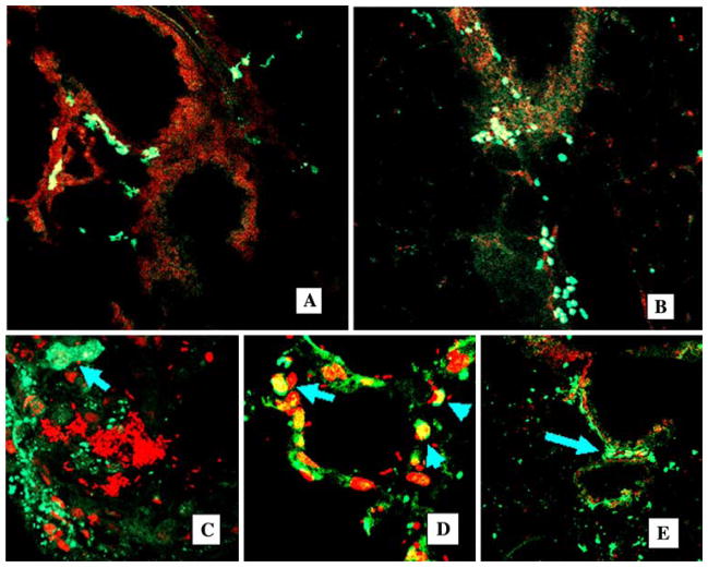
Fig. 2.
Chimerism in noninjured and injured lung. Confocal images of cryosectioned lung, nuclear stained with propidium iodine (red fluorescence). GFP+ cells are clearly visible as indicated by bright green fluorescence. A, B Lungs from mice after 1 month of 5.05-Gy irradiation and GFP+ BM cell transplantation (control group). Original magnification ×20. C–E Progeny of transplanted BMDC participate in acute lung inflammation. Confocal images of the lung, costaining PI (red), GFP+ cells are bright green (arrows). C After intratracheal administration of E. coli, lungs are infiltrated with GFP+ BMDC (bacterial pneumonia, 1 week). Abscess (bright red E. coli) is surrounded by GFP+ BMDC (arrow, original magnification ×40), costaining PI (red). D Multiple GFP+ BMDC are present in infected alveoli (original magnification ×40), costaining PI (red). E GFP+ cells in the peribronchial interstitium (arrows), original magnification ×20, costaining PI (red)

