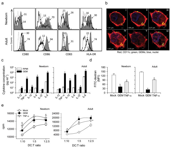
Figure 7.
GEM particles enhanced the maturation of neonatal and adult human DC and the stimulation of T cells in a MLR. (a) Expression of cell surface markers CD80, CD86, CD83 and HLA-DR on human CD11c+ neonatal and adult DC stimulated with GEM particles (shaded area) or mock- treated (solid line); the dashed line indicates the isotype control. The MFI on CD11c+ gated cells is indicated. (b) Individual (i–v) and Z-stack projection (vi) confocal laser microscopy images showing CD11c+ DC from human newborns harboring FITC-labeled GEM particles (arrows). CD11c+ cell-surface expression is shown in red, and nuclei are shown by blue fluorescent staining. Scale bars, 2 μm. (c) Cytokines produced by human neonatal CD34+- and adult derived DC stimulated with GEM particles or mock-stimulated and measured in culture supernatants. Data represent mean cytokine concentration±s.e.m. from two independent experiments. (d) FITC-dextran uptake by neonatal CD11c+ and adult human DC exposed to GEM or TNF-α (positive control), or mock-treated DC, measured by flow cytometry; data represents MFI±s.e.m. from three independent experiments. (e) Allogeneic stimulation of adult CD3+ T cells in the presence of neonatal and adult human DC stimulated with GEM particles or TNF-α, or mock-stimulated DC. Data show mean cpm±s.e.m. from one of two independent experiments. (c–e) Significant differences (*P<0.05) compared with mock-stimulated cells. GEM, Gram-positive Enhancer Matrix; MLR, mixed lymphocyte reaction, DC, dendritic cell; TNF, tumor necrosis factor; MFI, mean fluorescence intensity; HLA, human histocompatibility leukocyte antigen.

