FIGURE 1.
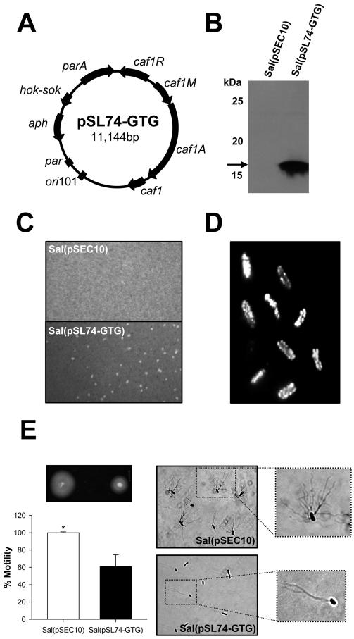
Expression of Y. pestis F1 by S. Typhi. A, Plasmid map of pSL74-GTG encoding the Y. pestis F1 operon. B, Western blot analysis of F1 expression. Bacterial lysates from S. Typhi strain ACAM948CVD carrying pSL74-GTG or empty plasmid pSEC10 were separated by SDS-PAGE. Proteins were transferred to polyvinylidene difluoride membranes, and F1 was revealed with F1-specific mAb. C, The formation of an external F1 capsule in S. Typhi(pSL74-GTG) was demonstrated by india ink staining; cultures were photographed under a light microscope with a Nikon Coolpix 4300 digital camera. D, Fluorescent microscopy confirming abundant F1 expression on the surface of S. Typhi(pSL74-GTG) upon incubation with F1-specific mAb and FITC-labeled anti-mouse IgG. No fluorescent staining was observed in S. Typhi(pSEC10) and S. Typhi(pSL74-GTG) incubated with normal mouse serum (data not shown). E,. Differential motility of S. Typhi(pSL74-GTG) and S. Typhi(pSEC10) and flagellar Ag expression shown by Ryu staining. *, p <0.05 indicates significant difference in motility between strains. Images were obtained with a ×100 objective.

