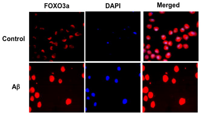
Figure 2.
During amyloid (Aβ1-42) exposure in inflammatory microglial cells, FoxO3a translocates to the cell nucleus to govern an initial activation and proliferation of microglial cells. Microglia were followed at 6 hours after Aβ1-42 (10 μM) (Aβ) administration with immunofluorescent staining for FoxO3a (Texas-red). Nuclei of microglia were counterstained with DAPI. In merged images, control cells have readily visible nuclei (white in color) that illustrate absence of FoxO3a in the nucleus. In contrast, merged images after Aβ1-42 (10 μM) exposure are not visible (red in color) and demonstrate translocation of FoxO3a to the nucleus. Control = untreated microglia.

