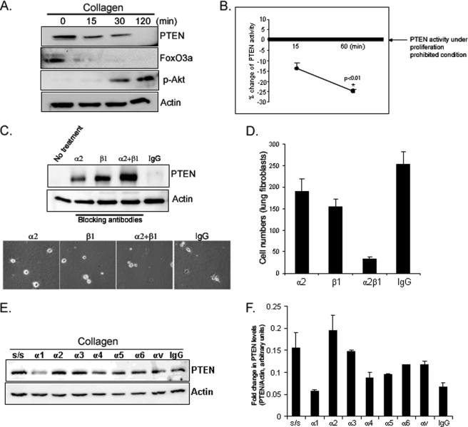
FIGURE 4.
Fibroblast adhesion to collagen decreases PTEN expression and activity. A, serum-starved fibroblasts were plated on dishes coated with type I collagen (100 μg/ml) and PTEN, FoxO3a, phosphorylated Akt (Ser-473), and actin levels were measured by Western analysis. B, serum-starved fibroblasts were plated on collagen for 15 and 60 min. p < 0.01 versus a reference value. Cells lysates were immunoprecipitated with anti-PTEN antibody, incubated with phosphatidylinositol (3,4,5)-trisphosphate substrate, and PTEN activity was quantified. To provide a reference value for PTEN activity under proliferation prohibitive conditions, PTEN activity was assessed in serum-starved fibroblasts cultured on tissue culture plastic plates under contact-inhibited conditions. C, upper panel, serum-starved fibroblasts were preincubated with α2- or β1-integrin blocking antibody (1 μg/ml), both (α2+β1), or isotype control antibody (IgG, 1 μg/ml) for 45 min and plated on collagen-coated plates for 30 min. PTEN levels were then measured. Actin was used as a loading control. Lower panel, shown are the phase-contrast microscopic cell morphologies at 30 min after plating the cells on collagen-coated plates. α2 and β1 represent α2 and β1 blocking antibody, respectively. IgG, isotype control. D, 5,000 cells were preincubated with integrin blocking antibodies for 45 min as indicated and the attached cells were counted at 30 min after plating the cells on collagen. E, serum-starved fibroblasts were pretreated with the indicated integrin blocking antibodies (1 μg/ml) and then plated on collagen-coated plates for 30 min. Total PTEN and actin levels were determined by Western analysis. As a reference, serum-starved (S/S) fibroblasts were trypsinized but not plated on collagen. F, PTEN expression was quantified by densitometry, normalized to actin.

