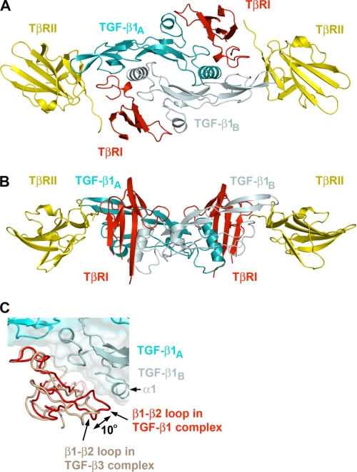
FIGURE 1.
Ribbon drawing of the TGF-β1 ternary complex. A, top view of the complex. B, side view rotated ∼90° compared with A. TGF-β1 monomers TGF-β1A and TGF-β1B are colored cyan and pale cyan, respectively. TβRI and TβRII are colored red and yellow, respectively. C, relative positions of type I receptors from TGF-β1 (red) and TGF-β3 (salmon) ternary complexes. This figure and all subsequent ribbon drawings are prepared using the PyMOL molecular graphics system.

