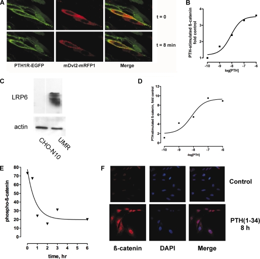
FIGURE 2.
Recruitment of Dvl and β-catenin activation, stabilization, and nuclear translocation following activation of PTH1R. A, PTH treatment induces translocation of Dvl2 to the plasma membrane. CHO cells expressing EGFP-PTH1R (green) were transiently transfected with mRFP-Dvl2. Confocal images were collected at 20-s intervals after the addition of 100 nm PTH(1–34). The images shown correspond to t = 0 and 8 min. Note the accumulation of red Dvl2 in discrete puncta (arrows, center panels). See supplemental Video 1. B, PTH-dependent β-catenin activation in CHO cells. CHO cells were transfected with HA-tagged human PTH1R and reporter TOP/FOP plasmids as described under “Experimental Procedures.” C, CHO cells do not express LRP6. The expression of LRP6 in UMR-106 cells is shown for comparative purposes. D, concentration-dependent stimulation of β-catenin activation by PTH. MC4 cells were transiently transfected with TOPFlash and 48 h later were studied as described under “Experimental Procedures.” E, stabilized β-catenin (i.e. dephosphorylated β-catenin) in MC4 bone cells after stimulation with PTH(1–34). F, translocation of activated β-catenin to the nucleus. ROS 17/2.8 cells were challenged with 100 nm PTH(1–34) for 8 h and then fixed and stained for dephosphorylated β-catenin and 4′,6-diamidino-2-phenylindole (DAPI), to stain nuclei.

