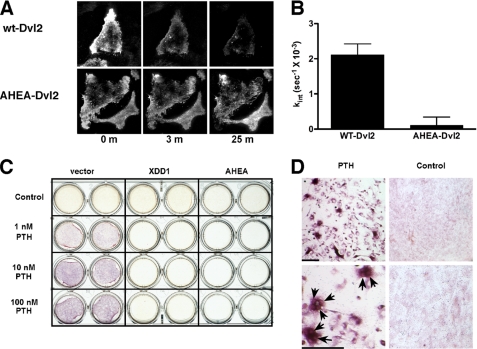
FIGURE 4.
Dvl is required for PTH-dependent PTH1R internalization and osteoclastogenesis. A, AHEA-Dvl2 blocks ligand-induced PTH1R internalization. Rat osteosarcoma cells transfected with PTH1R-EGFP and mRFP1-Dvl2 were imaged at 20-s intervals after the addition of 100 nm PTH(1–34) using TIRF. The data obtained with cells that express wild-type Dvl2 are shown in the top panel. The bottom panel shows the results of the expression of AHEA-Dvl2. B, summary of the PTH1R internalization results obtained. The rate constant of internalization of the PTH1R was calculated from TIRF microscopy data. Only cells that expressed both proteins (as determined from green and red fluorescence measurements) were used in these calculations. The data show the average of five different cell plates examined in three independent experiments. C, PTH promotes osteoclastogenesis through a β-catenin-mediated pathway. UAMS-32P cells were co-cultured with non-adherent bone marrow cells (52). PTH(1–34) at increasing concentrations was added in the presence of empty vector, XDD1 dominant negative Dvl, or AHEA dominant negative Dvl. After 6 days, tartrate-resistant acid phosphatase activity was determined using naphthol phosphate as a substrate and fast garnet to label the product as a red-purple precipitate. D, low and high magnification views of the cells shown in C. Tartrate-resistant acid phosphatase staining was primarily localized to multinucleated osteoclasts (arrowheads). Bar, 100 μm.

