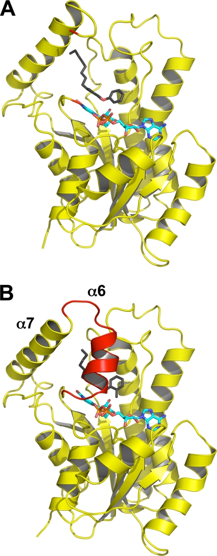
FIGURE 4.
Loop ordering upon slow binding inhibition. A, one monomer of the ternary InhA·NAD+·8PP complex (Protein Data Bank code 2b37) is shown in cartoon representation with the NAD+ molecule in cyan and the 8PP molecule in black and all-bonds representation. The substrate-binding loop is disordered in the 8PP structure, and the loop ends are depicted in red. B, monomer of the ternary InhA·NAD+·PT70 complex using the same colors and orientation as in A. The substrate-binding loop is ordered in this structure and covers the binding pocket (red cartoon). Secondary structure elements for both molecules were assigned with STRIDE (34).

