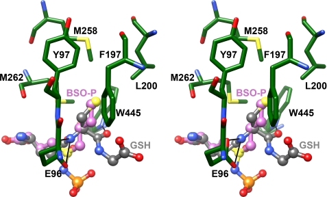
FIGURE 5.
Superpositioning of the glutathione and BSO binding sites indicate the location of the cysteine-binding site. Bound ADP and BSO are shown in ball and stick representation, and pertinent active site residues are shown in stick representation in the stereodiagram. Atoms are colored as in Fig. 2, with the exception of carbon atoms in BSO (colored in magenta). The S-butyl group of BSO and the thiol group of glutathione occupy a comparable hydrophobic pocket in the ScGCL active site.

