FIGURE 2.
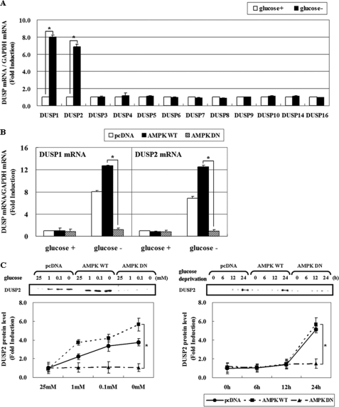
AMPK activation is critical for DUSP induction in response to glucose deprivation. A, HCT116p53+/+ cells were incubated in glucose-depleted medium for 24 h. Then, the mRNA levels of 12 DUSP members were determined by real-time PCR analysis, and the fold induction is expressed as a ratio of each DUSP: GAPDH mRNA. B, HCT116p53+/+ cells were transfected with pcDNA, pAMPK-WT, and pAMPK-DN expression vectors for 24 h, and maintained under glucose deprivation for 24 h. The amount of DUSP1 and -2 and GAPDH mRNA was evaluated by real-time PCR analysis. Results are the means ± S.E. for six determinations. C, HCT116p53+/+ cells were transfected with pcDNA, pAMPK-WT, and pAMPK-DN expression vectors for 24 h, and then were exposed to medium containing the indicated concentrations of glucose for 24 h (left panel) or incubated in glucose-free medium for the indicated times (right panel). The amount of DUSP2 protein was determined by Western blot analysis, and its level was quantified by densitometer. The data are expressed as the means ± S.E. (*, p < 0.05; compared with the indicated groups.) glucose+, 25 mm glucose; glucose-, 0 mm glucose.

