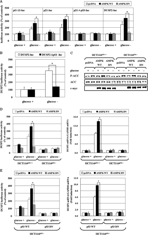
FIGURE 5.
AMPK increases DUSP2 promoter activity via p53 activation. A, HCT116p53+/+ cells were co-trasnfected with pcDNA, pAMPK-WT, or pAMPK-DN expression vectors, and a luciferase reporter containing a promoter with 13 p53 responsive elements (pG-13-luc), a wild-type p21cip1/waf1 promoter (p21-luc), p21 promoter with the p53 binding site deleted (p21cip1/waf1Δp53-luc), or pDUSP2-luc for 24 h. B, HCT116p53+/+ cells were transfected with a luciferase reporter with a wild-type DUSP2 promoter (pDUSP2-luc) or a DUSP2 promoter with the p53 binding site deleted (pDUSP2Δp53-luc) for 24 h. After exposure to glucose-depleted medium for 24 h, cell lysates were subjected to a luciferase activity assay. The data represent means ± S.E. for six determinations. C and D, HCT116p53+/+ and HCT116p53−/− cells were transiently co-transfected with pcDNA, pAMPK-WT, or pAMPK-DN expression vector, and pDUSP2-luc with a 1:1 ratio. After 24 h transfection, cells were exposed to glucose deprivation for 24 h, and then phosphorylation level of ACC and total ACC was measured by Western blot (C). Also, the luciferase activity assay (left panel) or mRNA level of DUSP2 was determined (right panel) (D). E, HCT116p53−/− cells were co-transfected with pDUSP2-luc and/or pcDNA, pAMPK-WT, pAMPK-DN, p53-WT, or p53-DN (p53-R175D) expression vectors at a 1:1:1 ratio. After 24 h transfection, cells were exposed to glucose deprivation for 24 h, and then luciferase activity (left panel) or mRNA level of DUSP2 (right panel) was measured. The data represent means ± S.E. for six determinations. (*, p < 0.05; compared with the indicated groups.)

