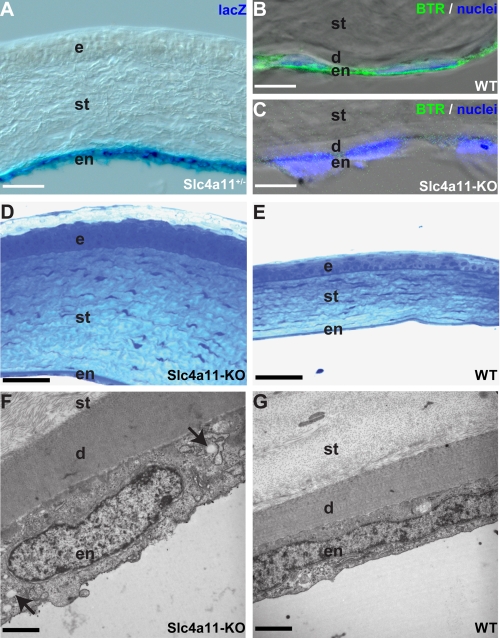
FIGURE 1.
Loss of SLC4A11 in endothelial cells leads to morphological alterations of the cornea. SLC4A11 is expressed in endothelial cells of the cornea (A and B). LacZ fusion protein expression (blue) in heterozygous animals (A) and antibody staining (green) in WT animals (B) reveals expression of SLC4A11 in the endothelium of the cornea. Antibody staining of KO tissue (C) proves specificity of the antibody. Nuclei in B and C are stained with TOTO-3 (blue). Sections through the cornea of slc4a11 knock-out (D) and WT mice (E) clearly reveal a significant thickening of the corneal stroma with disturbance of the stratification of the epithelium. Electron microscopy sections reveal a thickened Descement membrane and enlarged endothelial cells in slc4a11 knock-out (F) compared with WT mice (G). Note intracellular vacuolation (arrows in F) in the endothelial cells of KO animals. d, Descement's membrane; e, epithelium; en, endothelium; st, stroma. Scale bars used are as follows: A, 87 μm; B and C, 7 μm; D and E, 50 μm; F and G, 2 μm.

