FIGURE 3.
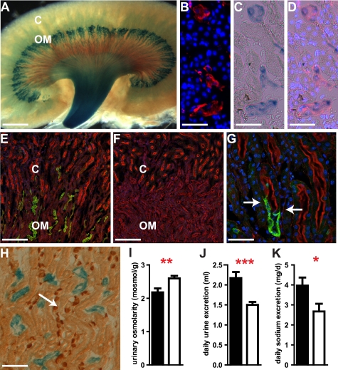
SLC4A11 in the thin descending limb of Henle loop is essential for urinary concentration. SLC4A11-LacZ fusion protein is localized (blue) in the medulla of the kidney (A). The expression of tubular aquaporin-1 (red, B) and of SLC4A11-LacZ (C, blue) co-localizes (D) in the medulla of the kidney. Nuclei are stained with TOTO-3 (blue; B, C, and G). SLC4A11 protein is detected in the medulla of the WT kidney (E and G; green), which coincides with the activity of SLC4A11-LacZ. SLC4A11 protein (green) is not present in proximal tubules (E and G) marked by staining of the brush border by phalloidin (red). SLC4A11 protein is absent in kidneys of KO animals (F). Localization of SLC4A11 protein (green) in the thin descending part of Henle loop close to the junction of the proximal tubule (marked by phalloidin staining in red) is shown in G (junction marked by arrows). Expression of SLC4A11-LacZ (blue) is not found in the collecting duct marked by AE1 staining (H; brown, arrow). Similarly, SLC4A11 is not detected in thick ascending limb and connecting tubule (see text). Loss of SLC4A11 leads to reduced urinary osmolarity (I) and increased daily urine (J) and sodium excretion (K) indicating defects in fluid resorption in the kidney (filled bars, mutants; open bars, WT; ***, p < 0.001; **, p < 0.01; *, p < 0.05; C, kidney cortex; OM, outer medulla; scale bars used are as follows: A, 1.2 mm; B and C, 43 μm; E and F, 174 μm; G, 43 μm; H, 100 μm).

