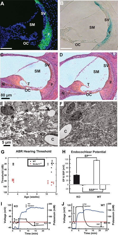
FIGURE 4.
Morphological alterations in the inner ear, impaired hearing, and loss of the endocochlear potential but preserved endolymph potassium concentration in SLC4A11 mutant mice. Anti-SLC4A11 antibody staining (green) in WT animals (A) reveals expression of SLC4A11 in the fibrocytes of the cochlea. Nuclei are stained with TOTO-3 (blue). LacZ fusion protein expression (blue) in heterozygous animals (B) confirms expression in fibrocytes. Hematoxylin and eosin-stained sections through the inner ear of slc4a11 knock-out (C) and WT mice (D) reveal no morphological changes at the light microscopy level. Electron microscopy images uncover abnormal cellular structures of fibrocytes in the stria vascularis of mutant (E) mice compared with WT (F), which are characterized by extracellular edema (arrowheads) and intracellular vacuolation (arrows). Measurement of hearing thresholds in KO and WT mice by auditory-evoked brain stem responses to clicks was recorded in anesthetized mice (G). Note the significantly reduced hearing threshold in KO mice at 3 weeks of age and in the older animals. H–J, strongly reduced endocochlear potential in KO mice compared with WT mice in the presence of normal potassium concentrations in the scala media. Representative measurements with combined EP (black line) and potassium recordings (blue line) are shown in I and J. The reference potential (red line) was recorded before the experiment in tissue (G1) and in the fluid meniscus (M1) overlying the cochlear opening. The reference potential was controlled at the same sites subsequent to the experiment (M2 and G2). Animals were sacrificed during endocochlear recording (T61 injection, Intervet), which causes an immediate loss of the endocochlear potential with subsequent formation of a passive steady state potential. C, capillaries; EP, endocochlear potential; F, fibrocytes; N, neurons; OC, organ of corti; SM, scala media; SSP, steady state potential; SV, stria vascularis; T, tectorial membrane; ***, p < 0.001.

