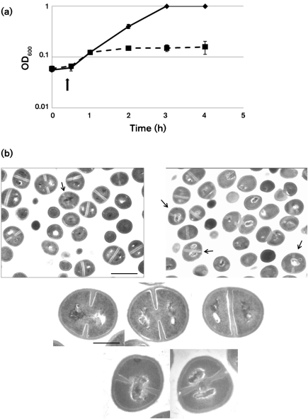
Fig. 5.
Effects of overexpression of Fst on S. aureus. (a) RNA Ipar was introduced into S. aureus UAMS-1 cells under control of the cadmium-inducible promoter of pCN51. Cadmium was added at 10 μM 30 min after culture dilution into antibiotic-free medium. Squares with broken line, pCN51 : : RNA Ipar; diamonds with unbroken line, pCN51. (b) Effects of the Fst toxin on cell morphology. The top row of panels shows a large field of induced pCN51 control (left) and pCN51 : : RNA I (right) cells to provide perspective on the proportion of abnormal cells. The arrow indicates a single cell with recognizable division septa and a centrally located nucleoid in the control panel. Note, however, that this cell is very early in the division process with only very small septal invaginations. In contrast, arrows in the right panel indicate two cells with centrally located nucleoids late in division and a third in which the nucleoid has been guillotined by the septum. Fst-exposed cells also occasionally displayed nucleoids pressed against the side of the cell, similar to those at the top of the image, probably due to completion of division in guillotined cells. Cells showing abnormal division septa, like the centrally located dividing cell, were more rarely seen. The middle row of panels shows the typical progression of nucleoid segregation seen in control cells, with the nucleoids showing some connection early in division (left), completely separating before the completion of division (middle), and well-separated by the time the septum is complete (right). The bottom row of panels shows close-ups of the two most common defects seen in Fst-exposed cells: centrally located nucleoids late in division (left) and guillotined nucleoids at the completion of division (right). Bars: top panels, 1 μm; middle and bottom panels, 0.3 μm.

