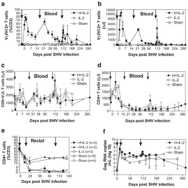
FIGURE 1.
HMBPP/IL-2 cotreatment initiated during early SHIV infection induces expansion of circulating Vγ2Vδ2 and αβ T cells but enhances viral infection. The relative percentage of CD3+ T cells that express Vγ2Vδ2 in the circulation (a) or CD4 in the rectal mucosa (e) and absolute numbers of circulating T cells per microliter of blood that express Vγ2Vδ2 (b), CD8+Vγ2− (c), or CD4+ (d) are shown over time for groups treated with HMBPP plus IL-2, IL-2 alone, or sham as averages ± SEM. Arrows indicate the time when treatment was given. Animals with severe prolonged CD4 T cell depletion (<3% of total CD3+ cells) in the rectal mucosa were stratified from the rest of their groups (e). Average SIVgag RNA copies per milliliter of plasma are shown over time for groups treated with HMBPP plus IL-2, IL-2 alone, or sham during both early and chronic infection (f). By Student’s t test, viral copy numbers are statistically higher (p < 0.05) at the following time points for HMBPP plus IL-2- (days 12, 22, 54, and 102) and IL-2 only- (days 12, 54, 102, and 123) treated groups compared with the sham-treated group (f). The viral levels substantially dropped at the last two data time points for the cotreated group since the two animals in this group with high viral loads did not survive at these time points.

