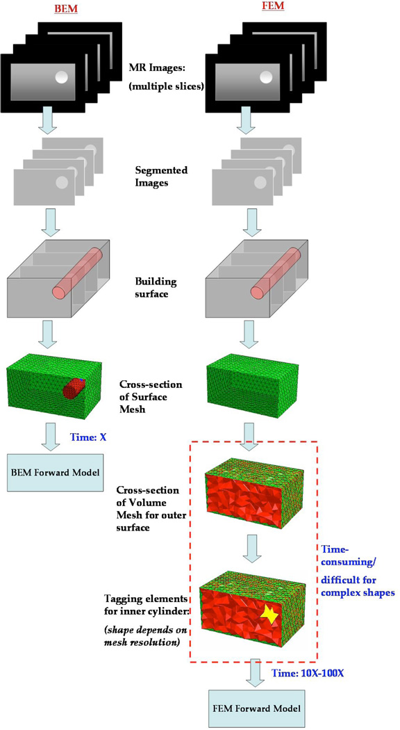
Fig. 1.
An illustration of the steps involved in obtaining a test grid for computation with BEM and FEM is shown. Both require segmentation of the MR dicom images followed by surface discretization. BEM can be used directly following the surface tessellation whereas FEM requires additional steps to build a volume mesh suitably tagged with boundaries delineating the volume into different tissue types. The shape of the interior boundaries will then depend on the mesh resolution. Alternately, the volume mesh can be created to preserve the interior boundaries (not shown here), however, this approach becomes difficult when resolving features at different scales (e.g. breast vs. tumor boundary). The volume meshing is a demanding task that takes up to 10–100 times the effort required to create surface meshes.

