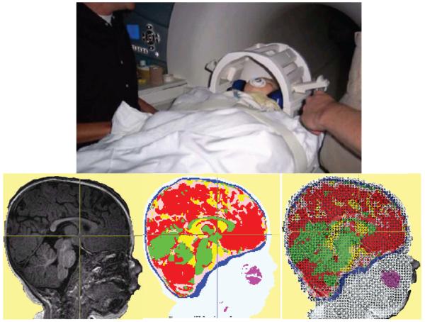
Fig. 2.
Structural magnetic resonance imaging (MRIs) for cortical source analysis of psychophysiological EEG (electroencephalogram) and ERP (event-related potentials). The top panel shows a 7.5-month-old (32 weeks) infant being placed into a Siemens 3T Trio Scanner. The procedure results in a MRI volume for the child's head, (bottom left), a MRI volume that segments the head materials (bottom center), and a high-resolution 3-D wireframe model representing the electrical model of the head used in computer programs for source analysis (bottom right).

