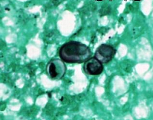
FIG. 11.
GMS-stained section of the lesion illustrated in Fig. 7 shows a budding yeast of B. dermatitidis with the characteristic broad-based bud, as well as two slightly misshapen single nonbudding yeast cells (high-power magnification). (Photograph courtesy of Jameel Ahmad Brown, University of Arkansas for Medical Sciences, Department of Pathology.)

