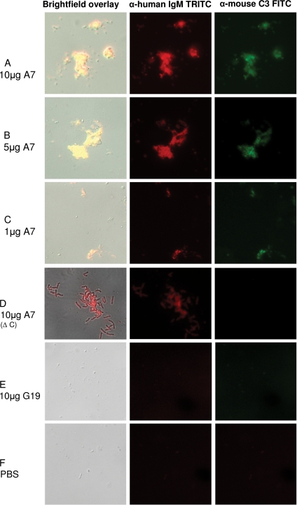
FIG. 5.
A7 mediates aggregation and complement deposition on ST3. Binding of IgM (red, middle) and C3 (green, right) and merged DIC bright-field images (overlay, left) are shown for the designated mixtures of ST3 (ATCC 6303), A7, and mouse serum (used as a complement source). Δ C, heat-inactivated complement source; G19, human IgM MAb to GXM. Magnification, ×100 (all images). The appearance of the A7-ST3 aggregate in panel D differs from that in panels A to C due to focusing on the aggregate, rather than the bacteria, in the bright-field image.

