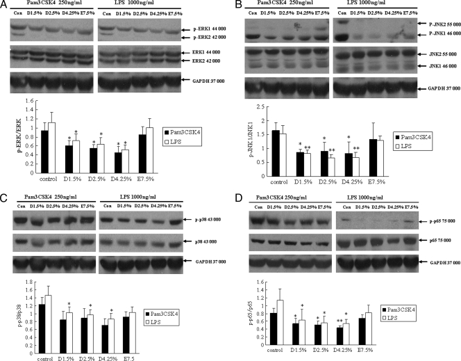
FIG. 3.
TLR ligand-induced MAPK and NF-κB signaling is reduced in glucose-based PD solution-treated human peritoneal mesothelial cells. Cells were pretreated with 1.5% Dianeal, 2.5% Dianeal, 4.25% Dianeal, and 7.5% icodextrin (Extraneal) for 48 h and then treated with TLR2 (Pam3CSK4; 250 ng/ml) or TLR4 (LPS; 1 μg/ml) ligand for 60 min. Immunoblot analysis was used to determine phosphorylated p38 MAPK, JNK, and p44/42 MAPK protein levels. Cells pretreated with glucose-based PD solutions that were stimulated with either Pam3CSK4 or LPS exhibited lower levels of phosphorylated p44/42 MAPK (A), JNK (B), p38 (C), and NF-κB p65 (D) than did untreated cells. Cells preincubated with icodextrin-based PD solutions did not shown an influence on TLR ligand-induced MAPK and NF-κB signaling. Data are expressed as means ± SD for three individual experiments. D1.5%, D2.5%, D4.25%, and E7.5% represent 1.5% Dianeal, 2.5% Dianeal, 4.25% Dianeal, and 7.5% icodextrin, respectively. *, P < 0.05; **, P < 0.01 versus control.

