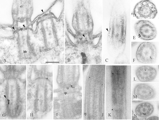
Fig. 4.
Localization of Bug22p in Paramecium by postembedding immunoelectron microscopy. (A to F) Labeling of wild-type cells with the GTL3 antibody using 5-nm gold particles. (G to N) Labeling of GFP-Bug22p-expressing cells with an anti-GFP antibody using 10-nm gold particles. The arrowheads point to gold particles. (A and H) Labeling of the terminal plates of basal bodies. (B, D, and I) Labeling of the transition zone to the cilium. (F and J to N) Labeling in the vicinity of the outer doublets of the axoneme. (C, E, and J) Labeling between the axoneme and the membrane. (G) Labeling within the basal body close to its proximal part. (J and K) Examples of regular disposition of the gold particles along the axoneme. ci, cilium; bb, basal body; tp, terminal plate; tz, transition zone. Bar = 250 nm.

