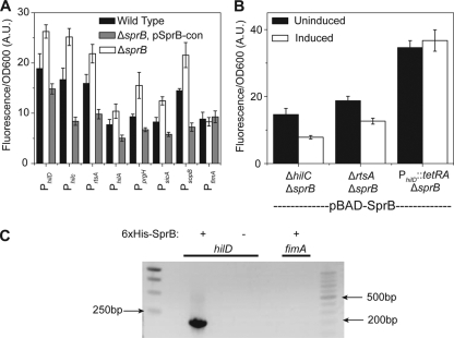
FIG. 3.
SprB represses SPI1 gene expression through HilD. (A) Comparison of SPI1 promoter activities in the wild type, a ΔsprB mutant, and a ΔsprB mutant constitutively expressing SprB from a plasmid (pSprB-con). (B) SprB repression of SPI1 gene expression is through the PhilD promoter. Comparison of PhilA promoter activity in ΔhilC ΔsprB, ΔhilC ΔsprB, and PhilD::tetRA ΔsprB mutants when SprB is expressed from an arabinose-inducible promoter on a plasmid (pBAD-SprB). SprB expression was induced with 2 mg/ml arabinose. In the experiments involving the PhilD::tetRA ΔsprB mutant, 2 μg/ml tetracycline was added to the growth medium in order to induce HilD expression. Induction in panel B is used to denote the presence or absence of arabinose. Fluorescence values were normalized with the optical density at 600 nm (OD600; A.U., arbitrary units) to account for cell density. Error bars indicate standard deviations. (C) SprB binds to the PhilD promoter region as determined by a coprecipitation assay using 6×His-tagged SprB. PCR was used to determine whether the PhilD promoter region is in the coprecipitated DNA. The PfimA promoter region was included as a negative control. For an expanded description of the experimental procedures, see the supplemental material.

