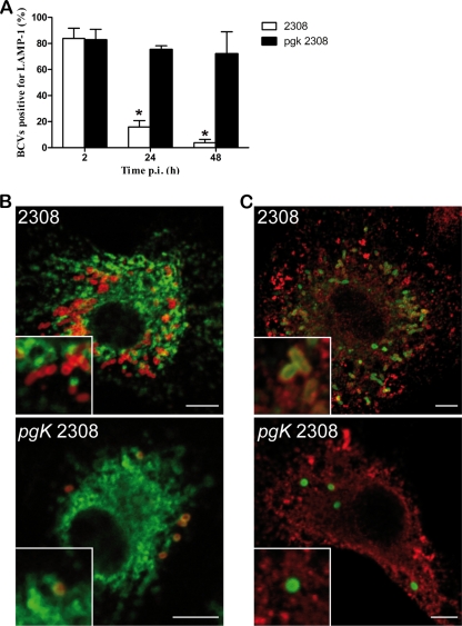
FIG. 4.
Multiplication and intracellular localization of B. abortus Δpgk mutant and wild-type strain in BMDM. (A) Quantification of the percentage of wild-type or Δpgk mutant BCVs that contain LAMP1 by confocal immunofluorescence microscopy. The difference between the wild type and mutant was statistically significant at 24 and 48 h (P < 0.001) postinfection (p.i.). Data are means from three different experiments. (B) Representative confocal images of BMDM at 24 h postinfection with wild-type B. abortus or the Δpgk mutant. Brucella lipopolysaccharide (LPS) is labeled in red, and LAMP1 is in green. (C) Confocal images of BMDM at 24 h postinfection with wild-type B. abortus or the Δpgk mutant. Brucella LPS is labeled in green, and calnexin is shown in red. Scale bar, 5 μm.

