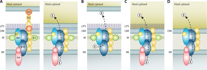
FIG. 5.
P-T4SS structural and functional diversification. The dashed-line arrows illustrate the mode of secretion, with substrates depicted by an encircled “s.” The color scheme of P-T4SS components VirB1 (green), VirB2 (light orange), VirB5 (dark orange), VirB7 (yellow), VirB9 (blue), VirB10 (dark blue), and VirD4 (pink) across four secretion systems implies homology. Distinct N and C termini of VirB10 and VirD4 are depicted. M, murein layer. (A) Model of transport for the A. tumefaciens vir P-T4SS. The two-step process of substrate attachment (left) and substrate transfer upon sloughing off of the T-pilus (right) is shown (51). (B) Model of transport for the B. pertussis ptl P-T4SS. VirB1 is distinguished to depict the N-terminal glycohydrolase domain of PtlE, a VirB8 homolog (107). (C) Model of transport for the Rickettsia rvh P-T4SS. (D) General model of transport for the O. tsutsugamushi rvh P-T4SS and for P-T4SSs in species of Anaplasmataceae that are not predicted to completely synthesize PG and LPS.

