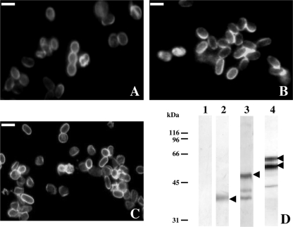
FIG. 1.
Immunolabeling in IFA and Western blotting with the monoclonal antibody 1E4. In IFA, MAb 1E4 reacts with the E. intestinalis spore wall (A). A cross-reaction is observed with the spore wall of both E. cuniculi (B) and E. hellem, strain EhD (C). (D) In the Western blot, a 40-kDa band, corresponding to the expected size of EiSWP1, is detected from E. intestinalis-infected HFF cells (lane 2). For E. cuniculi, a major band at 50 kDa (expected size for EcSWP1) is labeled (lane 3). The two bands at 40 and 42 kDa probably correspond to degradation products. For E. hellem, two major bands at 55 and 60 kDa are revealed (lane 4). Arrows indicate the major recognized proteins. Proteins were extracted from healthy and infected HFF cells in Laemmli buffer containing 2.5% SDS and 100 mM DTT and analyzed by 10% SDS-PAGE. Lane 1, healthy HFF cells; lanes 2, 3, and 4, HFF cells infected by E. intestinalis, E. cuniculi, and E. hellem (EhD), respectively. MAb 1E4 was diluted at 1:100 in IFA and 1:1,000 in Western blot. Bars, 1 μm.

