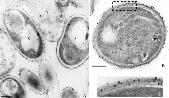
FIG. 5.
Immunoelectron microscopy of different E. hellem developmental stages with antibodies raised against the C-terminal part of SWP1b. In sporonts (A) and sporoblasts, a strong labeling is observed at the periphery of the cells. In spores (A and B), gold particles are mainly associated with the electron-dense outer layer of the spore wall (i.e., the exospore). Panel C corresponds to a higher magnification of the exospore area (boxed in panel B). The antisera were used at a 1:20 dilution. The secondary antibody was goat anti-mouse IgG conjugated with 10-nm colloidal gold particles (Sigma). SP, sporont; S, spore; PT, polar tube; Ex, exospore; En, endospore. Bars, 200 nm.

