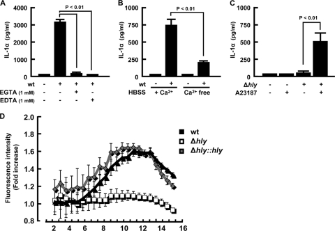
FIG. 6.
Involvement of intracellular calcium elevation in IL-1α secretion. Macrophages were infected with L. monocytogenes for 1 h, and EGTA or EDTA was added to the cultures at 3 h after infection (A). Alternatively, macrophages were infected with the wt for 1 h and cultured in calcium-free (Ca2+ free) or calcium-containing (+ Ca2+) RPMI1640 medium (B). The culture supernatants were collected at 18 h after infection, and IL-1α production was determined by ELISA. Macrophages were infected with the Δhly strain for 1 h and treated with 10 μM A23187 3 h later. The culture supernatant was collected 18 h after infection, and IL-1α production was measured (C). Macrophages were infected with the wt, Δhly, or Δhly::hly strain (MOI = 5) for 1 h, and the culture medium was replaced with dye-loading solution containing Fluo-4 NW at 2 h after infection. Following further incubation for 30 min, the fluorescence intensity was measured for 16 h at 30-min intervals. Results are expressed as fold increases in the fluorescence intensity over levels for nonstimulated controls. Data represent the averages and standard deviations of a quadruplet assay. Similar results were obtained in two independent experiments. HBSS, Hanks balanced salt solution.

