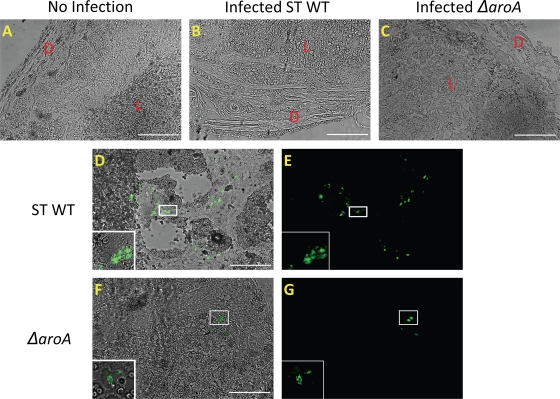
FIG. 9.
Localization of WT S. Typhimurium (ST) and S. Typhimurium ΔaroA bacteria to distinct regions of the placenta. Shown are images of frozen placental sections from mice infected with WT S. Typhimurium (103 CFU) or S. Typhimurium ΔaroA (106 CFU) expressing GFP 72 h postinfection. (A, B, and C) Morphology of the placentas from uninfected mice and mice infected with WT S. Typhimurium or S. Typhimurium ΔaroA, respectively. D, decidual region; L, labyrinth region. (D and E) Localization of WT S. Typhimurium-GFP in the highly necrotic labyrinth trophoblast. (F and G) Localization of S. Typhimurium ΔaroA-GFP near the decidual region of the placenta. The presence of bacteria was confirmed by observing the marked areas using a 60× oil immersion objective, and the higher-magnification images of bacteria are shown in insets. The images were captured using an Olympus IX81 fluorescence microscope. Scale bars, 200 μm. Images are representative of tissues processed from 3 mice per group.

