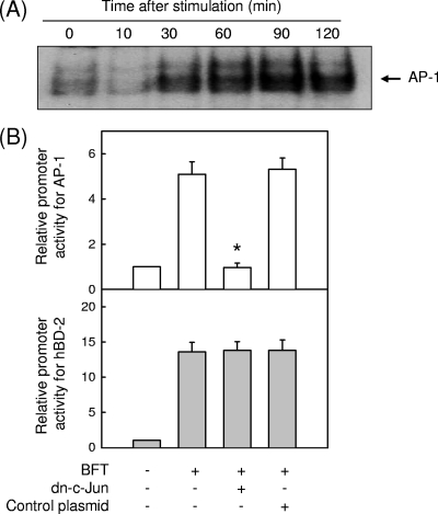
FIG. 4.
Relationship between AP-1 signaling and hBD-2 expression in BFT-stimulated HT-29 cells (A) HT-29 cells were stimulated with BFT (300 ng/ml) for the indicated periods of time. AP-1 activity was assessed by EMSA. The results are representative of three repeated experiments. (B) HT-29 cells were transfected with the pAP-1- or wild-type hBD-2-luciferase transcriptional reporter, together with the dominant-negative c-Jun superrepressor, as indicated. Then, 48 h later, the cells were stimulated with BFT (300 ng/ml) for another 1 h (AP-1) or 6 h (hBD-2), after which luciferase assays were performed. Data are expressed as mean fold induction in luciferase activity relative to unstimulated controls ± SEM (n = 5). The mean fold induction of the β-actin reporter gene relative to unstimulated controls remained relatively constant throughout each experiment. *, P < 0.05 compared with BFT alone.

