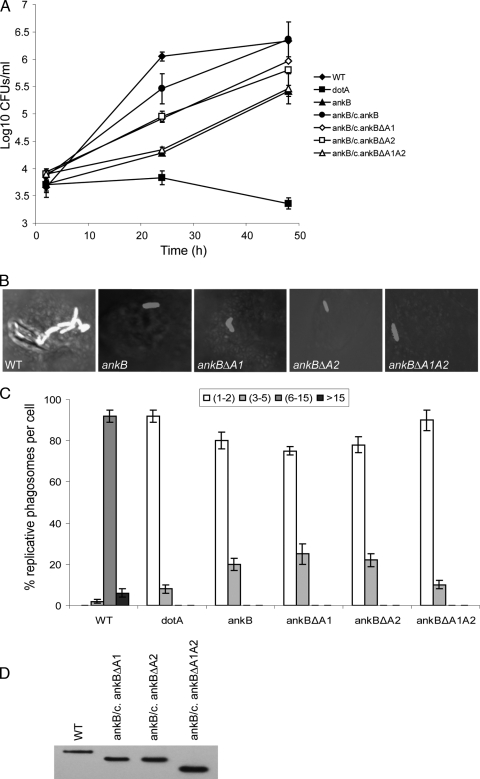
FIG. 1.
The two ANK domains of AnkB are essential for intracellular growth of L. pneumophila in macrophages. (A) Monolayers of U937 macrophages were infected with the WT strain and the isogenic dotA or ankB mutants or the ankB mutant complemented with either WT ankB (c/ankB) or one of the ankB mutant alleles. The infection was carried out in triplicate with an MOI of 10 for 1 h followed by 1 h of gentamicin treatment to kill extracellular bacteria. The infected monolayers were lysed at different time points and plated onto agar plates for colony enumeration. The results are representative of three independent experiments performed in triplicate. Error bars represent standard deviations. (B and C) Single cell analyses of L. pneumophila replicative phagosomes. At 10 h postinfection, 100 infected cells were analyzed by laser scanning confocal microscopy for formation of replicative phagosomes, and representative images are shown in panel B. L. pneumophila was stained by a polyclonal anti-L. pneumophila antibody and Alexa Fluor 555-conjugated anti-rabbit IgG (red). Quantification of the number of bacteria/cell at 10 h is shown in panel C. The dotA mutant was used as a negative control. Infected cells from multiple coverslips were examined in each experiment. The results are representative of three independent experiments performed in triplicate. Error bars represent standard deviations. (D) Immunoblot analysis shows no detectable differences in the expression levels of the AnkB variants in L. pneumophila. Total bacterial proteins equivalent to 1 × 10⁸ bacteria were loaded onto SDS-polyacrylamide gels and immunoblotted using an anti-AnkB rabbit antiserum (1:60,000 dilution).

