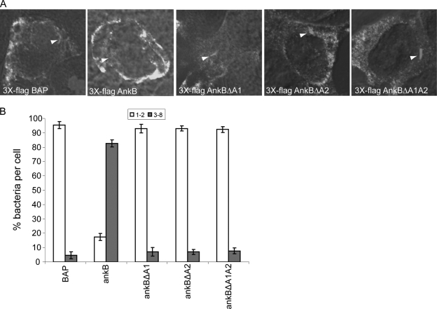
FIG. 8.
Ectopic expression of 3×Flag AnkBΔA1, AnkBΔA2, or AnkBΔA1A2 fails to rescue intravacuolar replication of the ankB mutant. HEK293 cells were transiently transfected with plasmids encoding 3×Flag BAP, 3×Flag AnkB, 3×Flag AnkBΔA1, 3×Flag AnkBΔA2, or 3×Flag AnkBΔA1A2 for 24 h prior to infection. Transfected HEK293 cells were then infected with the WT strain or the ankB or dotA mutant strain, and after 2 and 12 h, 100 infected cells were analyzed by confocal microscopy for formation of replicative phagosomes. (A) Representative confocal microscopy images of transfected HEK293 cells at 12 h postinfection. Ectopically expressed 3×Flag proteins were detected using antibody (green) while bacteria were detected using a rabbit anti-L. pneumophila antibody (red). Nuclei were stained with DAPI (blue). White arrowheads indicate bacteria inside cells. (B) Quantitation of single cell analysis of ankB mutant replicative phagosomes in transfected HEK293 cells 12 h postinfection. At 2 h and 12 h post-infection of HEK293 cells, 100 infected cells were analyzed by confocal microscopy for formation of replicative phagosomes. The WT strain and the dotA mutant bacteria were used as positive and negative controls, respectively. Infected cells from multiple coverslips were examined in each experiment. All the results are representative of three independent experiments performed in triplicate. Error bars represent standard deviations.

