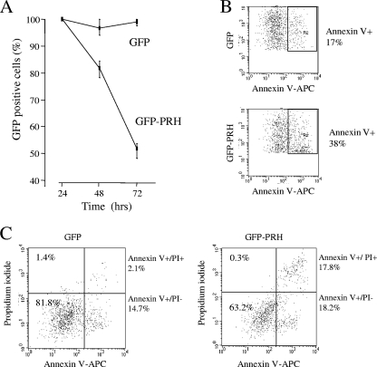
FIG. 5.
PRH overexpression induces apoptosis in K562 cells. (A) Cells transfected with pEGFP alone or pEGFP and pMUG1-Myc-PRH. The percentage of GFP-expressing cells 24 h posttransfection was set as 100%, and the change from this was tracked over 72 h. Values are means and SD (n = 3). (B) Cells from panel A were analyzed by flow cytometry. The percentage of GFP-positive/annexin V (AV)-positive cells was determined 24 h posttransfection. One representative dot plot of 3 is shown. (C) Cells from panel A were dual stained with propidium iodide (PI)/AV (APC antibody) and analyzed by flow cytometry. The dot plot shows the percentages of live cells (PI− AV−), necrotic cells (PI+), early apoptotic cells (AV+), and late apoptotic cells (AV+ PI+) after gating for GFP+ cells. One representative dot plot of 3 is shown.

