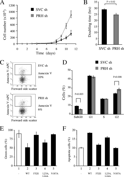
FIG. 6.
PRH KD increases cell proliferation. (A) Cumulative growth curves for K562 cells transfected with SVC shRNA (sh) or PRH shRNA (1+2). Cells were selected with puromycin 24 h posttransfection. After 7 days, 3 × 10⁶ cells of each cell type were plated out and counted over 11 days. (B) Graph of the doubling time for K562 cells transfected with SVC shRNA or PRH shRNA (1+2) (gray bar). Values are means and SD (n = 3). (C) Cells from panel A were dual stained with PI/AV (APC antibody) and analyzed by flow cytometry. The dot plots show percentages of live cells (AV−) and percentages of apoptotic cells (AV+). (D) Percent distribution of cells in each stage of the cell cycle. PI staining of K562 cells transfected with SVC shRNA or PRH shRNA (1+2) is shown (n = 3). (E) Percentages of GFP+ cells in cotransfection experiments. K562 cells were transfected with pEGFP and either pMUG1 (bar 1) or pMUG1 vectors expressing PRH (bar 2; WT, wild type), PRH F32E (bar 3), PRH L23A,L24A (bar 4), or PRH N187A (bar 5), and the percentage of GFP+ cells was measured 72 h posttransfection. Values are means and SD (n = 3). (F) K562 cells were transfected as for panel E and dual stained with PI/AV antibody 24 h posttransfection for analysis by flow cytometry (n = 3).

