FIG. 7.
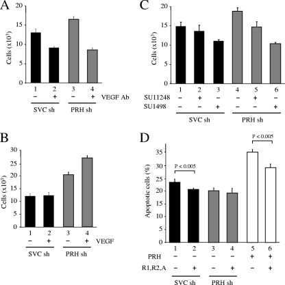
PRH KD cells are sensitive to VEGF inhibition. (A) K562 cells were transfected with SVC shRNA (sh) or PRH shRNA and then incubated with 50 μg/ml anti-VEGF antibody (bars 2 and 4) or an equal volume of DMSO (bars 1 and 3) for 72 h. An MTT assay was then used to calculate cell numbers. Values are means and SD (n = 5). (B) As for panel A except that cells were incubated with 50 ng/ml VEGF. Values are means and SD (n = 5). (C) As for panel A except that cells were incubated with DMSO (bars 1 and 4), 2 μM SU11248 (bars 2 and 5), or 2 μM SU1498 (bars 3 and 6). Values are means and SD (n = 3). (D) K562 cells transfected with SVC shRNA (black bars) or PRH shRNA (gray bars) or not shRNA transfected (white bars) were then transfected with a vector expressing PRH (3 μg) (bars 5 and 6) and/or vectors expressing VEGF, VEGFR-1, and VEGFR-2 (bars 2, 4, and 6) (total, 3 μg). Twenty-four hours posttransfection the cells were dual stained with PI/AV antibody for analysis by flow cytometry. The graph shows the means and SD (n = 3).

